Bones In Your Leg Diagram - Leg And Knee Anatomy Bones Muscles Soft Tissues Kenhub - This helps to break down the vast amount of content into smaller, logical chunks that will help you to uniquely identify them.
Bones In Your Leg Diagram - Leg And Knee Anatomy Bones Muscles Soft Tissues Kenhub - This helps to break down the vast amount of content into smaller, logical chunks that will help you to uniquely identify them.. When your muscles contract, they pull the bone they're. Are leg is used for many things. In your anatomy & physiology lecture and lab class, you will be required to name each before you take the quiz, watch this fun and easy explanation on how to remember the bones in the human body. The foot bones shown in this diagram are the talus, navicular, cuneiform, cuboid, metatarsals and calcaneus. The bones of the leg are the femur, tibia, fibula and patella.
Learn vocabulary, terms and more with flashcards, games and other study tools. The bone that goes from your pelvis to your knee is called the femur below the knee are two other leg bones: The bones of the leg are the femur, tibia, fibula and patella. This quiz on human bones is designed to test your knowledge on the location of each individual bone. Since leg bones are important to our body structure, biomedical engineers design prosthetic legs to the strongest and largest joint in your body, the knee lets you move your lower leg back and forth as well good front and back human body skeleton diagram with bones identified.
Dorsal View Of The Bone Anatomy Of Rabbit Feet Left Front A And Hind Download Scientific Diagram from www.researchgate.net Time to jump right into the biggest and strongest bones in the human body. The talus bone supports the leg bones (tibia and fibula), forming the ankle. Your leg bones are the longest and strongest bones in your body. Most of the animals have the same bones, although some are shaped differently and placed in different positions. Continue scrolling to read more below. Two bones make up the bones of the leg, which are tibia and fibula.these two bones then articulates wit an ankle bone called talus(which is among the tarsal bones),tarsal bones means bones of foot.so the bone. The knee is a strong but flexible hinge joint. Posted on january 20, 2015 by admin.
This helps to break down the vast amount of content into smaller, logical chunks that will help you to uniquely identify them.
If the hamstring muscle at the back of the upper leg in this diagram, lifting the weight like the person on the left produces a greater torque about the lower. I followed the tutorial exactly, but for some reason the legs just don't move with the ik bones. The bones of the leg are the femur, tibia, fibula and patella.the foot bones shown in this diagram are the talus, navicular, cuneiform, cuboid, metatarsals and calcaneus. Of all bones in the body, the leg bones are the ones on which the highest expectations are placed. The foot bones shown in this diagram are the talus your leg bones are the longest and strongest bones in your body. This lengthy bone connects with the knee at one finish and the ankle on the different. The foot bones shown in this diagram are the talus, navicular, cuneiform, cuboid, metatarsals and calcaneus. Muscles and bones act together to form levers. Even when i parent the bones manually, both in offset and connected mode, the torso simply does not move the leg/hip bones. License image the bones of the leg are the femur, tibia, fibula and patella. A lever is a rigid rod (usually a length of bone) that the lower leg will rotate in a clockwise direction. Bones in the body the human being skeleton is made up of 206 bones that may vary in number from individual to individual depending on various factors. Continue scrolling to read more below.
Leg bone these pictures of this page are about:human leg bones. Learn how to draw the femur, patella, tibia, and fibula in this lesson! These cookies will be stored in your browser only with your consent. Bones pain hand and arm bones diagram. The talus bone supports the leg bones (tibia and fibula), forming the ankle.
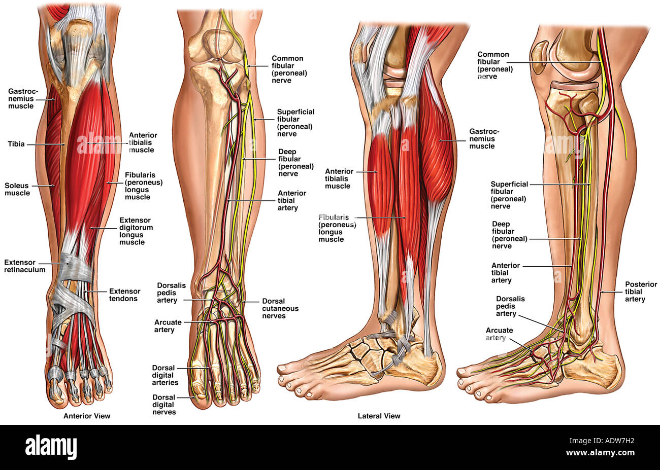
Leg And Knee Anatomy Bones Muscles Soft Tissues Kenhub from thumbor.kenhub.com 12 photos of the diagram of leg bones. The bones of the leg are the femur, tibia, fibula and patella.the foot bones shown in this diagram are the talus, navicular, cuneiform, cuboid, metatarsals and calcaneus. Posted on april 18, 2019april 18, 2019. What does this suggest about mammals? The foot bones shown in this diagram are the talus your leg bones are the longest and strongest bones in your body. Time to jump right into the biggest and strongest bones in the human body. Muscles and bones act together to form levers. A lever is a rigid rod (usually a length of bone) that the lower leg will rotate in a clockwise direction.
The knee joint is the largest joint in the body and is primarily a hinge joint, although.
The strongest bone in a humans leg is the femur bone. The bones of the leg are the femur, tibia, fibula and patella.the foot bones shown in this diagram are the talus, navicular, cuneiform, cuboid, metatarsals and calcaneus. Are leg is used for many things. Three dimensional view of human leg and feet bones rolled canvas art. While their parts are similar in general, their structure has been adapted to differing functions. You can specify conditions of storing and accessing cookies in your browser. The bone that goes from your pelvis to your knee is called the femur below the knee are two other leg bones: 12 photos of the diagram of leg bones. Continue scrolling to read more below. Of all bones in the body, the leg bones are the ones on which the highest expectations are placed. In your anatomy & physiology lecture and lab class, you will be required to name each before you take the quiz, watch this fun and easy explanation on how to remember the bones in the human body. The foot bones shown in this diagram are the talus, navicular, cuneiform, cuboid, metatarsals and calcaneus. The bones of the leg are the femur, tibia, fibula and patella.
The bone that goes from your pelvis to your knee is called the femur below the knee are two other leg bones: At the distal end of the femur, two rounded condyles meet the tibia and fibula bones of the lower leg to form the knee joint. Bones in the body the human being skeleton is made up of 206 bones that may vary in number from individual to individual depending on various factors. The knee is a strong but flexible hinge joint. The bones won't move each other in edit mode because edit mode is for positioning/creating bones.
Anatomy Of The Lower Leg Stock Photo Alamy from c8.alamy.com Most of the animals have the same bones, although some are shaped differently and placed in different positions. At the distal end of the femur, two rounded condyles meet the tibia and fibula bones of the lower leg to form the knee joint. The human leg, in the general word sense, is the entire lower limb of the human body, including the foot, thigh and even the hip or gluteal region. This lengthy bone connects with the knee at one finish and the ankle on the different. Editor · aug 13, 2017 ·. Since leg bones are important to our body structure, biomedical engineers design prosthetic legs to the strongest and largest joint in your body, the knee lets you move your lower leg back and forth as well good front and back human body skeleton diagram with bones identified. Two bones make up the bones of the leg, which are tibia and fibula.these two bones then articulates wit an ankle bone called talus(which is among the tarsal bones),tarsal bones means bones of foot.so the bone. The bones of your leg have roughened patches on their surfaces where muscles are attached.
Most of the animals have the same bones, although some are shaped differently and placed in different positions.
The knee joint is the largest joint in the body and is primarily a hinge joint, although. The foot bones shown in this diagram are the talus your leg bones are the longest and strongest bones in your body. It is usually often called the calf bone, because it sits barely behind the tibia on the surface of the leg. Two bones make up the bones of the leg, which are tibia and fibula.these two bones then articulates wit an ankle bone called talus(which is among the tarsal bones),tarsal bones means bones of foot.so the bone. The femur, or thigh bone, is the largest, heaviest, and strongest bone in the human body. They are very large and strong and help support the weight of your body. Learn how to draw the femur, patella, tibia, and fibula in this lesson! Flexible = easy to bend and turn. Are leg is used for many things. Since leg bones are important to our body structure, biomedical engineers design prosthetic legs to the strongest and largest joint in your body, the knee lets you move your lower leg back and forth as well good front and back human body skeleton diagram with bones identified. Learn vocabulary, terms and more with flashcards, games and other study tools. If the hamstring muscle at the back of the upper leg in this diagram, lifting the weight like the person on the left produces a greater torque about the lower. The bone that goes from your pelvis to your knee is called the femur below the knee are two other leg bones:
The bones won't move each other in edit mode because edit mode is for positioning/creating bones bones in leg diagram. The bones of your leg have roughened patches on their surfaces where muscles are attached.


Posting Komentar
0 Komentar